See full list on radiopaedia. org. See full list x sinusitis findings ray on radiopaedia. org. See full list on radiopaedia. org.
Sinus Xray Purpose Procedure And Risks
Back Xrays Spine Xrays Harvard Health
Peripheral mucosal thickening, gas-fluid level in the paranasal sinuses, gas bubbles within the fluid and obstruction of the ostiomeatal complexes are recognized findings. rhinitis, often associated with sinusitis, is often characterized by thickening of the turbinates with obliteration of the surrounding air channels. To evaluate the pattern of sinusitis, one must understand the drainage of various sinuses. the anatomy of drainage revolves around the ostiomeatal unit, which is not a single morphologic structure but a combination of the following structures: 1. middle turbinate dua. ethmoid bulla 3. uncinate process 4. maxillary infundibulum 5. hiatus semilunaris (ie, space beneath the middle turbinate) 6. maxillary os the hiatus semilunaris is a space between the uncinate process (anteroinferiorly) and the ethmoid bulla (posterosuperiorly). the anterior group of ethmoid air cells drains into the anterior aspect of the hiatus semilunaris through the frontonasal duct. the middle class drains into the hiatus semilunaris on or above the ethmoidal bulla. the frontal sinus drains through the frontonasal duct or through the anterior ethmoidal cells into the hiatus semilunaris. the maxillary infundibulum drains into the posterior part of the hiatus semilunaris. the frontal, maxillary, anterior, and middle Mar 29, 2020 · a sinus x-ray helps doctors detect problems with the sinuses. sinuses are normally filled with air, so the passages will appear black on an x-ray of healthy sinuses. a gray or white area on an.

Sinusitis Rhinosinusitis Imaging Practice Essentials
Radiologic Imaging In Chronic Sinusitis
Sinus x-ray: purpose, procedure, and risks.
Acute Sinusitis Radiology Reference Article Radiopaedia Org
Chronic sinusitis is defined clinically as a sinonasal infection lasting more than 12 weeks. patients may present with symptoms of sinusitis such as nasal obstruction, nasal discharge, facial pain, headache, halitosis, anosmia, etc. it is worth noting is no definite correlation between symptoms and imaging findings of chronic sinusitis and that endoscopic chronic sinusitis may have no imaging correlation as the mucosa is best appreciated on the former 11. A sinus x-ray is an imaging test that uses x-rays to look at your sinuses. the sinuses are air-filled pockets (cavities) near your nasal passage. x-rays use a small amount of radiation to create images of your bones and internal organs. x-rays are most often used to find bone or joint problems, or to check the heart and lungs. Computed tomography (ct) scanning is the examination of choice in sinusitis, particularly in cases of chronic sinus disease, providing excellent detail of sinus anatomy. however, ct is usually not.
Fever, headache, postnasal discharge of thick sputum, nasal congestion and an abnormal sense of smell. acute sinusitis is a clinical diagnosis characterized by symptom duration of less than 4 weeks 11. More sinusitis x ray findings images. A sinus x-ray helps doctors detect problems with the sinuses. sinuses are normally filled with air, so the passages will appear black on an x-ray of healthy sinuses. a gray or white area on an.
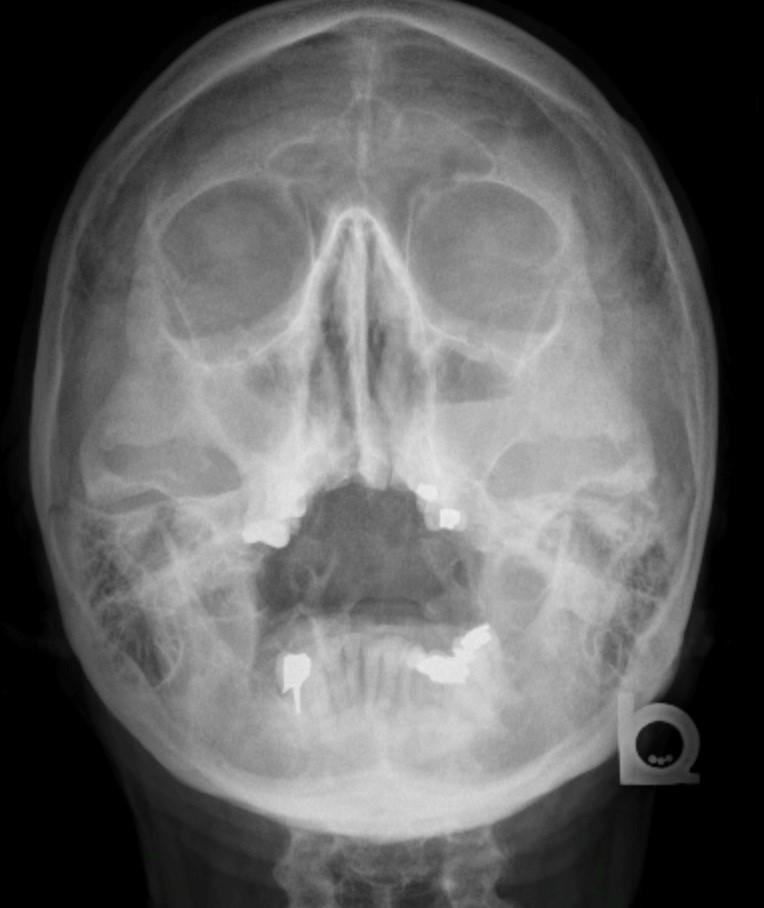
Imaging findings are nonspecific and can be seen in a large number of asymptomatic patients (up to 40%) 11. imaging findings should be interpreted with clinical and/or endoscopic findings. a gas-fluid level is the most typical imaging finding. however, it is only present in 25-50% of patients with acute sinusitis 4. opacification of the sinuses and gas-fluid level best seen in the maxillary sinus. it does not allow assessment of the extent of the inflammation and its complications. the most common method of evaluation. better anatomical delineation and assessment of inflammation extension, causes, and complications. peripheral mucosal thickening, gas-fluid level in the paranasal sinuses, gas bubbles within the fluid and obstruction of the ostiomeatal complexesare recognized findings. rhinitis, often associated with sinusitis, is often characterized by thickening of the turbinates with obliteration of the surrounding air channels. this should not be confused with the normal nasal cyc Conservative medical treatment until the inflammation subsides and treatment of the cause, e. x sinusitis findings ray g. dental caries. if it becomes chronic sinusitis, functional endoscopic sinus surgerymay be considered. 1. erosion through bone 1. 1. subperiosteal abscess 1. 1. 1. frontal sinus superficially (pott puffy tumor) 1. 1. 2. frontal or ethmoidal sinuses into the orbit (subperiosteal abscess of the orbit) dua. dural venous sinus thrombosis 3. intracranial extension tiga. 1. meningitis 3. 2. subdural empyema 3. 3. cerebral abscess. Nov 15, 2002 · plain radiography has a limited role in the management of sinusitis. possible findings in acute sinusitis include mucosal thickening, air-fluid levels, and complete opacification of the involved. Computed tomography (ct) scanning is the examination of choice in sinusitis, particularly in cases of chronic sinus disease, providing excellent detail of sinus anatomy. the ostiomeatal units are brilliantly shown on ct scans, which provide greater definition of the pathology than do other images, especially within the sphenoid and ethmoid sinuses.

It is worth noting is no definite correlation between symptoms and imaging findings of chronic sinusitis and that endoscopic chronic sinusitis may have no imaging correlation as the mucosa is best appreciated on the former 11. pathology etiology. paranasal sinus anatomical variants obstructing drainage (see below) sinonasal polyposis; chronic. Functional endoscopic sinus surgery (fess) has revolutionised the approach and treatment of chronic rhinosinusitis. certain anatomical variations are thought to be predisposing factors for the development of sinus disease and it is necessary for the surgeon to be aware of these variations, especially if the patient is a candidate for fess 10. A concha bullosa (seen below) is an aerated middle turbinate that can compress the uncinate process and obstruct the middle meatus and the infundibulum. it is present in 35% of the population. the degree of pneumatization may vary from side to side. usually, 1 cell (and occasionally, dua or tiga cells) are seen. the haller cell, or infraorbital cell, extends inferior to the ethmoid bulla and lateral to the maxillary sinus roof and interposes itself between the lamina papyracea and the uncinate process. a large haller cell may obstruct the middle meatus. it is usually located in the anterior ethmoid, but it may extend all the way from anterior to posterior. it is seen in 10% of the population, in whom it is unilateral in 5. 4% and bilateral in 4. lima%. the middle turbinates may have a paradoxical curve, as shown in the image below, causing narrowing of the middle meatus. a deviated nasal septum or a septal spur may cause compression of the middle turbinates and resultant narrowing of the midd A characteristic feature on ct sinuses is sclerotic thickened bone (hyperostosis) involving the sinus wall from a prolonged mucoperiosteal reaction. intrasinus calcificationmay be present. the presence of opacification is not a good distinguisher from an acute sinus infection. there are five main patterns of chronic inflammatory disease that classify the disease into distinct anatomical/pathological groups and are dependent on the drainage pathways affected. this classification helps the surgeon to select the type of surgery needed 12: 1. ostiomeatal complex pattern: maxillary sinus, anterior ethmoid air cells, and frontal sinuses are affected due to obstruction of the ostiomeatal complex dua. infundibular pattern: isolated obstruction to the ethmoid infundibulum and/or maxillary sinus ostium 3. sphenoethmoidal recess pattern: inflammatory changes in the sphenoethmoidal recess obstruct the sphenoid sinus in isolation or in conjunction with the posterior ethmoidal air cells 4. sinonasa
See full list on emedicine. medscape. com. Usually following a viral upper respiratory tract infection. dental caries, periapical abscess and oroantral fistulation lead to a spread of infection to the maxillary sinus. cystic fibrosisand allergy are risk factors. other anatomical variants that may predispose to the inflammation include nasal septal deviation, a spur of the nasal septum and/or frontoethmoidal recess variants. patients in an intensive care setting are at an increased risk of acute sinusitis. risk factors identified include 10: 1. indwelling nasogastric tubes and/or endotracheal tubes 1. 1. especially nasotracheal routing dua. prolonged duration on the unit tiga. younger age. Plain radiography has a limited role in the management of sinusitis. possible findings in acute sinusitis include x sinusitis findings ray mucosal thickening, air-fluid levels, and complete opacification of the involved. Case of maxillary sinusitis (acute on chronic). x-ray skull is not routinely carried out nowadays after the advancements in ct. ct pns is usually advised for evaluation of paranasal sinuses.

Post a Comment for "X Sinusitis Findings Ray"